Affected person cohort
This retrospective examine included sufferers who underwent routine contrast-enhanced MRI examinations of the liver or prostate as a part of their customary medical care between March 2022 and April 2024. The examine was accredited by the Mass Common Brigham Institutional Assessment Board (IRB). All strategies had been carried out in accordance with the related institutional tips and laws. As a result of retrospective nature of the examine, the Mass Common Brigham IRB waived the requirement for knowledgeable consent. The next exclusion standards had been utilized: age < 18 years, hepatospecific contrast-enhanced research, incomplete examinations, scanning of a number of physique areas in a single examination (together with MRCP), and incomplete information extraction from the scanner log. After making use of these standards, we excluded a complete of 650 examinations: 52 exams as a consequence of age < 18 years, 45 exams for hepatospecific contrast-enhanced research, 58 exams for incomplete examinations, 473 exams for scanning a number of physique areas (together with MRCP), and 22 exams as a consequence of incomplete information extraction from the scanner log. This left 2,723 MRI examinations included within the closing evaluation.
MRI services
All examinations had been acquired on 3T MRI scanners (MAGNETOM Vida, Siemens Healthineers, Forchheim, Germany) at two belly MRI services inside our hospital community: one established outpatient reference facility and a brand new, optimized MRI facility. The RF is a extremely environment friendly, multi-modality imaging heart that employs a conventional MRI scanner setup with a single, fastened scanning desk and one preparation bay. Adjoining to the scanner suite, a delegated space for peripheral intravenous line placement by nursing workers allows streamlined affected person preparation. Nonetheless, this association required sequential affected person processing, with the subsequent affected person ready till the present examination was accomplished earlier than coming into the scanner room for preparation. In distinction, the OF, which started operations in March 2022, is an unique MRI facility that includes an progressive structure designed to reinforce effectivity and affected person throughput. It contains three MRI scanners and three devoted preparation bays. Every scanner is supplied with a pair of interchangeable dockable tables with a corresponding duplicate set of coils. The affected person preparation bays are positioned in a direct path to the scanner, separated by a central hall and are used for affected person positioning, intravenous catheter placement, coil and earplugs/headphone placement, permitting for concurrent affected person preparation and scanning. As soon as the previous examination was accomplished, the tables had been swapped instantly, enabling a seamless transition between sufferers and minimizing congestion. Moreover, the MRI scanners are every angulated to facilitate a quicker docking course of. An in depth floorplan of the brand new imaging facility was revealed beforehand by Lang et al.10. This progressive design allowed for a discount in slot instances from 45 min at RF to 30 min at OF.
Each services maintained standardized staffing ranges with two MRI technologists per scanner. The optimized facility moreover employed one technologist aide shared throughout all three scanners to help the parallel workflow enabled by the preparation bay and dockable desk system. The aide assisted with affected person transport and preparation duties, permitting technologists to give attention to scanning procedures.
Picture acquisition
Picture acquisition at each services was carried out utilizing similar protocols carried out throughout establishments. The liver protocol consisted of Dixon T1-weighted imaging yielding in/opposed-phase/fats/water pictures, T2-weighted imaging with and with out fats suppression and diffusion-weighted imaging with corresponding ADC maps. Dynamic contrast-enhanced imaging was obtained after administration of gadoterate meglumine (0.1 mmol/kg, Dotarem®; Guerbet, Princeton, NJ, USA) at 1–2 mL/s adopted by a saline flush, buying pre-contrast, arterial (35–40 s), portal venous (60–75 s), and delayed section (3–5 min) pictures.
The prostate protocol consisted of high-resolution T2-weighted imaging in axial, sagittal, and coronal planes, diffusion-weighted imaging with corresponding ADC maps, and dynamic contrast-enhanced imaging following intravenous administration of gadobutrol (Gadavist, Bayer HealthCare) at a dose of 0.1 mmol/kg physique weight at a price of two mL/s, adopted by a 20 mL saline flush.
The reference facility began incorporating deep studying (DL) reconstruction methods in late 2021, whereas the optimized facility utilized DL reconstruction from its inception in March 2022.
Information extraction and workflow metrics
Our evaluation relied on two main information sources: automated scanner logs supplied by the seller (Siemens Healthineers, Forchheim, Germany), which contained timestamps for all MRI scanner occasions, and digital well being information (EHR) examination information. These datasets had been precisely matched by examination IDs and overlapping timestamps.
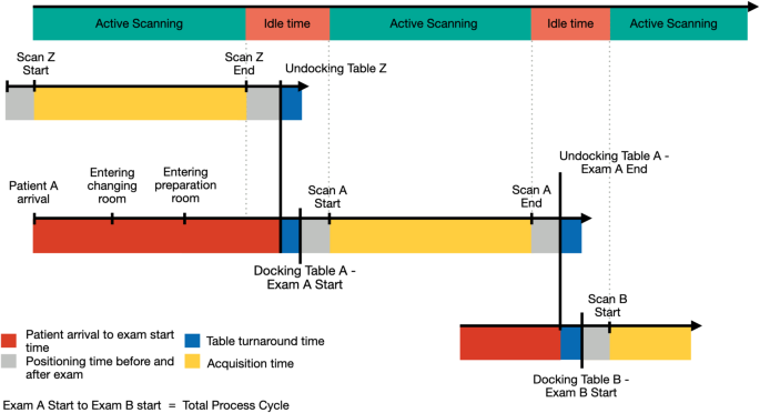
From this mixed dataset, we extracted and evaluated a number of key workflow metrics (Fig. 1). The affected person arrival to examination begin time measured the interval between a affected person’s check-in on the imaging facility and the beginning of their examination. We additionally assessed the entire course of cycle, which encompassed the complete period from the beginning of 1 examination to the start of the subsequent. This cycle comprised three distinct parts: positioning time, acquisition time, and desk turnaround time. Positioning time displays the important affected person positioning and preparation steps that happen on the scanner desk itself within the scanner room. It’s outlined because the sum of two particular intervals: (1) the time between when the scanner first detects desk exercise (corresponding to vertical desk motion or docking) till the beginning of the primary imaging sequence, and (2) the time between the top of the ultimate imaging sequence till the scanner’s final detection of desk exercise (corresponding to undocking or desk motion). Acquisition time represented the period of lively picture acquisition, measured from the beginning of the primary sequence to the top of the final. Desk turnaround time captured the interval between the final occasion related to one affected person and the primary occasion of the next affected person on the identical scanner (i.e. vertical desk motion, un-/docking desk). To make sure information integrity and account for affected person availability, we solely calculated desk turnaround time when the subsequent affected person arrived no less than 10 min earlier than the conclusion of the present examination.
Schematic diagram outlining the phases of the MRI examination course of. The highest row shows the scanner standing, alternating between lively scanning (inexperienced) and idle time (coral). The three timelines beneath reveal a sequence of affected person examinations. The center timeline follows Affected person A’s full examination cycle: affected person arrival to examination begin time (pink) begins when Affected person A enters the ability and contains time in altering room and preparation bay; desk turnaround time (blue) is outlined because the interval between undocking Affected person Z’s desk and docking Affected person A’s desk; positioning time (grey) represents each the preliminary interval between desk docking and begin of scanning, and the ultimate interval between final sequence and desk undocking; acquisition time (yellow) captures the period of lively scanning. The underside timeline exhibits the start of Affected person B’s examination as Affected person A’s concludes. The whole course of cycle is outlined because the interval from the beginning of 1 examination to the beginning of the subsequent (e.g., Examination A Begin to Examination B Begin).
Whereas the scanner log information supplied information for course of cycle time, acquisition time, positioning time, and desk turnaround time, we extracted affected person arrival and scheduled examination instances from the EHR.
Statistical evaluation
All calculations had been carried out utilizing SPSS (Model 29.0.0.0, IBM Corp., Armonk, NY), and all graphics had been generated utilizing R Studio (Model 2024.04.2 + 764 Posit, PBC, Boston, MA). Descriptive statistics had been performed for affected person traits and examination distribution throughout the imaging services and over time.
A 3-way evaluation of variance (ANOVA) was used to evaluate the impression of facility design, physique area, and date (impartial variables) on the important thing workflow metrics extracted from the scanner log (dependent variables). Estimated marginal means (EMM) and customary errors (SE), in addition to the mode for desk turnaround time, had been reported. Submit-hoc evaluation utilizing Bonferroni correction was performed for pairwise a number of comparisons.
On-time efficiency was calculated for every facility kind to match the proportion of examinations beginning inside 5 min of the scheduled time. Chi-square exams had been used to evaluate variations in categorical variables between the 2 services.
A 95% confidence interval was used, and p-values < 0.05 had been thought-about statistically vital.
Declaration of AI and AI-assisted applied sciences within the writing course of
Through the preparation of this work the authors used “Claude 3.5 Sonnet AI Assistant. Anthropic, PBC “in an effort to enhance the readability, language and high quality of the writing. After utilizing this service, the authors reviewed and edited the content material as wanted and take full duty for the content material of the publication.


